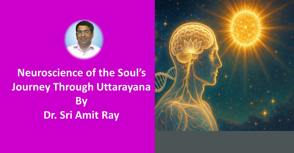
Benefits and Neuroscience of Ek-Sruti Mantra Chanting

    Mantra chanting is a powerful spiritual practice that has been used for millennia across various cultures and traditions. It harnesses the power of sound to influence the mind, body, and spirit. Among the various forms of mantra chanting, Ek-Sruti (monotone chanting) stands out due to its simplicity and effectiveness. Ek-Sruti chanting involves the repetition of a mantra on a single, unchanging pitch, creating a continuous and steady sound vibration.

    In this article, we explore the profound benefits of Ek-Sruti (monotone chanting) mantra chanting and delve into its neuroscience, demonstrating how this practice affects brain function, mental states, and overall well-being.

    "Ek-Sruti mantra chanting is the voice of stillness, where the mind dissolves into the resonance of a single note, unlocking the subtle energies of the soul and the cosmos." - Sri Amit Ray

    Ek-Sruti mantra chanting combined with the beeja mantras of the Ray 114 chakras offers a unique and powerful path for spiritual awakening, emotional healing, and mental clarity. The steady sound of the monotone pitch aligns with the specific frequencies of each chakra, facilitating the activation, cleansing, and balance of these energy centers.

    Ek-sruti chanting is different from melodic chanting. Melodic chanting also known as Sāma-gāna, incorporates melody and rhythm into the chanting process. The tones may rise and fall, following specific scales or ragas, which makes the chanting more musical in nature. 

    Benefits of Ek-Sruti Monotone Mantra Chanting

    This type of mantra chanting, often used in meditation or minimalist compositions, may affect brainwave patterns, emotional states, and cognitive processing in several ways:

    1. Brainwave Synchronization 

    Monotone chanting, especially with rhythmic repetition, can lead to brainwave synchronization, where the brain’s electrical activity syncs with the external auditory stimulus. This often induces specific states such as:

    • Alpha waves (8-12 Hz): Associated with relaxation and meditative states. Repetitive, monotone music can promote alpha wave production, leading to a state of calm and mental clarity. Studies found that mantra chanting  leads to increase in alpha relative power. Increase in relative alpha band power is associated with the secretion of hormones or neurotransmitters causing dilation of blood vessels which is further associated with the improvement in the mental well-being and feeling relaxed and releived, thus leading to decrease in anxiety and depression [1]. 
    • Theta waves (4-8 Hz): Related to deep relaxation, creativity, and dreamlike states. This can occur during prolonged exposure to low-frequency, monotonic sounds, often used in meditative practices.

    2. Reduction in Cognitive Load

    Monotone mantra chanting lacks the complexity of more dynamic musical compositions, which can reduce cognitive load. This reduction allows the brain to enter a more passive or relaxed state, potentially lowering stress and anxiety. This effect is useful in practices such as Om meditation or 114 chakras meditation, where the goal is to quiet the mind and achieve a state of inner calm.

    3. Activation of the Default Mode Network (DMN)

    Repetitive and predictable sound patterns, like monotone music, can deactivate attention-demanding networks in the brain and allow for the activation of the default mode network (DMN). The DMN is associated with mind-wandering, daydreaming, and self-referential thoughts. Listening to monotone music may encourage introspective thinking and self-awareness [2].

    4. Impact on the Emotional Centers of the Brain

    Monotone music can evoke a wide range of emotional responses, often dependent on the context or listener’s mental state. Since it lacks traditional musical dynamics (melody, harmony), the listener’s emotional response is less about the music's complexity and more about their internal interpretation. Some possible effects include:

    • Calming or meditative effect: The repetitive nature can create a hypnotic or soothing atmosphere.
    • Boredom or frustration: For some, the lack of variety can be perceived negatively, leading to feelings of discomfort.

    5. Auditory Cortex and Minimalist Perception

    The brain’s auditory cortex processes the sound of monotone music differently than more varied musical compositions. The simplicity of the music allows for the auditory system to focus less on processing changes in pitch, harmony, or rhythm, and more on other auditory features like timbre or texture. This can enhance mindful listening and awareness of subtler sonic elements.

    6. Therapeutic Uses

    Monotone music has been used in therapeutic settings, especially in mindfulness and meditation, where it helps facilitate mental stillness. It is also employed in sound therapy to promote relaxation and reduce symptoms of anxiety or depression by inducing specific neural states.

    7. Cognitive Flexibility and Long-Term Effects

    Listening to monotone music over time may lead to a more adaptable or flexible cognitive state. Since the music doesn’t demand active engagement or analysis, it can allow the brain to disengage from external distractions, promoting a sense of inner focus or flow. This may help with creative problem-solving or reflective thinking.

    Brain Areas and Mantra Chanting

    Read more ..


    Neuroscience of Samadhi: Brainwaves, Neuroplasticity, and Deep Meditation

    The neuroscience of Samadhi, a state of deep meditative absorption and heightened awareness, offers intriguing insights into how the brain and nervous system function during advanced meditative states.

    Samadhi, especially as described in the Yoga Sutras of Patanjali, represents a union of mind, body, and consciousness. The study of these states from a scientific perspective involves looking at brainwave patterns, neuroplasticity, brain regions involved, and biochemical changes. Let's explore the key aspects:

    This article explores the intersection of neuroscience and deep spirituality, focusing on Samadhi's impact on brainwaves, neuroplasticity, and the transformative effects of deep meditation. It examines how these elements contribute to profound states of consciousness and cognitive spirituality for total wellbeing

    What is Samadhi

    In yogic philosophy, Samadhi is defined as the highest state of mental absorption, where the practitioner experiences complete unity with the object of meditation, and eventually, with consciousness itself. It is the final stage of the eight limbs of yoga, leading to Kaivalya—liberation from the cycle of birth and death. 

    There are two major categories of traditional Samadhi:

    1. Samprajnata Samadhi (with cognition): In this state, the mind still operates with a focus on subtle objects like thought, emotions, or abstract ideas. There is awareness, but it is absorbed in deep concentration on finer aspects of reality.
    2. Asamprajnata Samadhi (without cognition): Also known as Nirbija Samadhi (seedless), this is a state of complete cessation of thought and perception. It is beyond the mind and ego, resulting in a profound sense of unity and non-dual awareness.

    Compassion and Samadhi 

    In our Ray 114 Chakras tradition, compassion and Samadhi are deeply interconnected, reflecting a profound synthesis of spiritual practice and inner transformation.

    Compassion, as a fundamental aspect of spiritual evolution, aligns with the higher vibrations of the 114 chakras, each representing different facets of the self and the universe. Samadhi, the pinnacle of meditative absorption, facilitates a state where individual consciousness merges with universal consciousness, fostering an expansive sense of empathy and interconnectedness.

    In this state, the meditator experiences a boundless compassion that transcends personal limitations, resonating with the pure essence of divine love and cosmic harmony. This synergy between compassion and Samadhi not only enhances personal spiritual growth but also contributes to the collective well-being, manifesting a holistic approach to spiritual enlightenment and human connection.

    Neuroscientific Definition of Samadhi

    Samadhi is a highly advanced state of neurocognitive integration characterized by profound, sustained neural synchronization and coherence across various brain regions, leading to a unified experience of consciousness. The samadhi state involves a shift from the default mode network's typical activity to a dominant activation of the brain's attentional and sensory processing networks, resulting in an enhanced perception of unity, reduced sense of ego, awakening of a heightened sense of present-moment awareness, and deep compassion. It reflects a profound transformation in neural processing, where the individual experiences a deep sense of inner stillness and integration, transcending ordinary cognitive and emotional states.

    Spiritual Definition of Samadhi

    Samadhi is the ultimate state of meditative absorption where individual consciousness seamlessly merges with universal consciousness, leading to a profound awakening of total cosmic well-being. It represents the pinnacle of spiritual realization, characterized by a profound sense of oneness and unity with all existence.

    In the state of samadhi, the meditator transcends the ego and dualistic perceptions, experiencing an all-encompassing bliss, compassion, creativity, and a deep ineffable peace. Samadhi is marked by the dissolution of personal desires and mental fluctuations, leading to a direct, experiential realization of one's true nature and the divine essence underlying the universal compassion. It is both a profound inner awakening and a complete surrender to the transcendent, unchanging reality.

    Types of samadhi in patanjali yoga sutras

    In Patanjali's Yoga Sutras, Samadhi is the final stage of meditation and is described in several types. Here’s a list of the key types:

    1. Samprajñāta Samadhi:

    • Savitarka Samadhi: This is a form of Samadhi where the meditator is focused on a gross object or concept, with active discernment and logical reasoning.
    • Savichara Samadhi: This type involves subtle, more refined objects of meditation, where the discernment is more abstract and subtle compared to Savitarka.
    • Sananda Samadhi: In this state, the meditator experiences bliss and joy, where the focus is on the pure essence of joy rather than external objects.
    • Sasmita Samadhi: This involves the sense of ego or individuality in a refined manner, where the meditator recognizes the self in its purest form.

    2. Asamprajñāta Samadhi:

    • Nirvikalpa Samadhi: A higher state of Samadhi where there is no differentiation or vikalpa (conceptualization); the meditator experiences pure consciousness without any mental modifications.
    • Nirbija Samadhi: The final stage of Asamprajñāta Samadhi, where there is no seed of desire or thought left, leading to complete liberation and transcendence.

    These types of Samadhi represent different levels of meditative absorption and realization, ranging from the more conceptual to the most profound state of pure consciousness.

    Neuroscience of Samadhi

    1. Brainwave Activity in Samadhi

    Meditative states associated with Samadhi are reflected in unique brainwave patterns:

    • Gamma Waves (30-100 Hz): These are associated with heightened cognitive function, concentration, and a state of “oneness” with the environment. Studies on advanced meditators show increased gamma activity, particularly in regions related to attention and sensory processing. In deep Samadhi, this heightened focus without an object reflects non-dual awareness.
    • Theta Waves (4-8 Hz): Associated with deep relaxation, creativity, and meditation, theta waves are prominent in meditative absorption. These waves often dominate when the mind is deeply quiet, introspective, and approaching the stillness that characterizes Samadhi.
    • Alpha Waves (8-12 Hz): These waves reflect a relaxed but alert state, often seen in light meditation. Alpha waves dominate early stages of meditation and gradually give way to deeper states as one moves toward Samadhi.

    In Samprajnata Samadhi (where awareness of objects or subtle mental content remains), alpha and theta waves may be more prominent, while Asamprajnata Samadhi (where awareness of all content disappears) may exhibit more gamma synchronization, indicating deep integration and unity of awareness.

    2. Neuroplasticity and Long-term Brain Changes

    Samadhi can induce profound long-term changes in the brain, often referred to as neuroplasticity:

    • Increased cortical thickness: Long-term meditators often show greater thickness in areas of the brain responsible for attention, sensory awareness, and emotional regulation, particularly the prefrontal cortex and insular cortex [1].
    • Reduction in the size of the amygdala: The amygdala, responsible for fear and stress responses, becomes less active and physically smaller with sustained meditative practice. This reflects a reduced reactivity to stress, fear, and negative emotions—common outcomes in advanced meditative states like Samadhi [2].
    • Increased connectivity in the default mode network (DMN): The DMN, which is active during self-referential thoughts and daydreaming, tends to quiet down during deep meditation. In Samadhi, the DMN is significantly suppressed, indicating a reduction in ego-centered activity and an experience of "oneness" or ego dissolution [3].

    3. Key Brain Regions Activated in Samadhi

    Several brain regions have been found to play a crucial role during Samadhi and advanced meditative states:

    • Prefrontal Cortex: This region is associated with higher-order thinking, attention, and self-regulation. During Samadhi, the prefrontal cortex shows increased activation, reflecting heightened concentration, and awareness, which is needed to sustain deep meditative states.
    • Parietal Lobe: This region processes sensory input and spatial awareness. Studies on experienced meditators suggest that the parietal lobe becomes less active during deep states like Samadhi, reducing the sense of separation between the self and the external world. This contributes to the feeling of "unity consciousness."
    • Insula: Involved in body awareness and interoception (awareness of internal bodily states), the insula is activated during meditation, including Samadhi. This may contribute to the sense of heightened awareness of the body’s energy, breath, and subtle sensations.
    • Anterior Cingulate Cortex (ACC): The ACC is associated with attention control, emotional regulation, and error detection. In Samadhi, the ACC is highly active, reflecting the capacity to maintain prolonged focus without distraction.
    • Thalamus: The thalamus acts as a relay station for sensory information. During Samadhi, thalamic activity is often altered, resulting in the filtering out of unnecessary external sensory input, allowing the practitioner to maintain deep meditative absorption.

    4. The Role of Neurotransmitters and Hormones

    During states of Samadhi, specific neurotransmitters and hormones play a role in the experience of bliss, focus, and calm:

    • Dopamine: Increased levels of dopamine, a neurotransmitter associated with reward and pleasure, are observed during meditation. This may explain the feelings of deep contentment and bliss often reported during Samadhi.
    • Serotonin: Known for its role in mood regulation, serotonin levels also rise during meditation, contributing to a sense of inner peace and well-being.
    • GABA (Gamma-aminobutyric acid): Meditation has been shown to increase GABA levels, a neurotransmitter that reduces neuronal excitability and anxiety. This calming effect may be part of the deep relaxation and tranquility experienced in Samadhi.
    • Endorphins: These natural painkillers and mood elevators are often released during meditation, leading to feelings of euphoria and detachment from physical sensations in advanced meditative states.

    5. Integration of the Sympathetic and Parasympathetic Nervous Systems

    Meditative practices leading to Samadhi involve the balancing of the autonomic nervous system:

    • Sympathetic Nervous System (SNS): Normally associated with fight-or-flight responses, the SNS becomes less active during deep meditation. Stress levels decrease, as evidenced by lower cortisol levels (a stress hormone).
    • Parasympathetic Nervous System (PNS): The PNS, responsible for rest and digestion, becomes dominant in Samadhi. This leads to slower heart rate, reduced blood pressure, and a state of deep physiological rest.

    6. Cognitive and Emotional Benefits of Samadhi

    • Enhanced Emotional Regulation: Meditators who reach Samadhi often experience profound emotional regulation. This is because regions such as the amygdala (fear and stress response center) and the prefrontal cortex (rational decision-making) work in harmony, reducing reactivity to external stimuli.
    • Increased Focus and Cognitive Function: The sustained attention required to enter Samadhi results in improved cognitive functions, including memory, attention, and decision-making.
    • Reduction of Egoic Thought: With the reduction of activity in the default mode network, egoic thinking diminishes. This allows practitioners to experience a sense of "selflessness," contributing to feelings of unity and interconnectedness.

    7. Samadhi as a State of Flow

    From a psychological perspective, Samadhi can be compared to the flow state:

    • Flow state: A state in which individuals are fully immersed in an activity, with a sense of energized focus, full involvement, and enjoyment. Samadhi, however, is deeper and more sustained than typical flow states, as it extends beyond engagement with tasks into a state of pure awareness without objectification.

    8. Long-term Psychological Effects of Samadhi

    The long-term attainment of Samadhi can have profound psychological effects:

    • Resilience and Emotional Intelligence: Regular meditation leading to Samadhi increases the brain’s capacity to handle stress, improve emotional intelligence, and develop resilience.
    • Bliss and Compassion: Advanced practitioners often report heightened feelings of compassion, joy, and a deep sense of love for all beings. This could be attributed to the combination of neurochemical changes and the silencing of egoic mental activity.

    Conclusion: Neuroscience Meets Samadhi

    The neuroscience of Samadhi reveals the profound physiological, cognitive, and emotional transformations that occur during advanced meditation. Through altered brainwave patterns, structural changes in the brain, and shifts in neurotransmitter levels, the brain reflects the stillness, clarity, and bliss associated with Samadhi.

    These changes not only align with spiritual descriptions but also suggest that Samadhi represents a harmonious state of optimal functioning in the human brain, bridging ancient spiritual wisdom with modern scientific understanding.

    References:

    1. Ray, Amit. The Science of 114 Chakras in Human Body: A Guidebook. Inner Light Publishers, 2015.
    2. Ray, Amit. "Epigenetic Reprogramming for Reversal of Aging and to Increase Life Expectancy." Amit Ray, amitray. com 2.4 (2023): 81-83, https://amitray.com/epigenetic-reprogramming-for-reversal-of-aging/
    3. Ray, Amit. "Slow Breathing Yoga Pranayama to Reduce Oxidative Stress." Compassionate AI, 1.3 (2024): 15-17. https://amitray.com/slow-breathing-yoga-pranayam-to-reduce-oxidative-stress/
    4. Ray, Amit. “Hormones, Endocrine System, and Your Seven Chakras: Balancing Your Body Mind and Spirit.” Amit Ray, September 27, 2023. https://amitray.com/hormones-endocrine-system-and-your-seven-chakras/
    5. Ray, Amit. “Neuroscience of Samadhi: Brainwaves, Neuroplasticity, and Deep Meditation.” Amit Ray, September 16, 2024. https://amitray.com/neuroscience-of-samadhi/.
    6. Ray, Amit. “Heart Rate Variability with Om Meditation and Chanting.” Amit Ray, August 8, 2024. https://amitray.com/stress-relief-and-heart-rate-variability-with-om-meditation/.
    7. Ray, Amit. "Neuroscience of Samadhi: Brainwaves, Neuroplasticity, and Deep Meditation." Compassionate AI, 3.9 (2024): 48-50. https://amitray.com/neuroscience-of-samadhi/
    Read more ..

    Slow Breathing Yoga Pranayama to Reduce Oxidative Stress

    Oxygen, the elixir of life, is indispensable for our existence, playing a pivotal role in cellular respiration and energy production. However, recent scientific observations have illuminated a paradox: while oxygen is vital for life, excessive oxygen intake can lead to oxidative stress [1], a condition associated with various diseases.

    "With harmony and peace in every inhale and exhale, yoga pranayama whispers the art of reducing oxidative stress and profound well-being." - Sri Amit Ray

    Slow breathing

    Slow breathing

    This realization has prompted a closer examination of ancient breathing practices, particularly resistance pranayama, as a potential remedy for mitigating oxidative stress. Researchers observed that relaxation induced by diaphragmatic breathing boosts the body's antioxidant defense system [2] of the body.

    In this article we explore the intricate relationship between oxygen, oxidative stress, and yoga slow breathing exercises. We explore the power of yoga slow breathing exercises, and their benefits for modern health.

    Recent breathing research has shown that quick, shallow and unfocused breathing may contribute to a host of problems, including anxiety, depression and high blood pressure. However, by harmonizing the equilibrium of oxygen and other respiratory gases, slow breathing exercises in yoga pranayama may contribute to diminishing oxidative stress and fostering overall well-being.

    Oxidative Stress and Diseases

    Research has established a strong correlation between oxidative stress and the pathogenesis of various diseases. Reactive oxygen species (ROS) function as crucial signaling molecules, intricately involved in the advancement of inflammatory disorders. Extensive research has underscored the significant correlation between oxidative stress and the development of various diseases.

    Even a modest elevation in lung vascular pressure has been shown to activate pro-inflammatory responses and increase ROS production in endothelial cells [3]. This imbalance between ROS production and antioxidant defenses is implicated in the development of conditions such as cancer, asthma, and pulmonary hypertension.

    Pranayama and Heart rate variability (HRV)

    Heart rate variability (HRV) refers to the variation in the time interval between heartbeats. It is considered a marker of the balance between the sympathetic nervous system (which governs the body's "fight or flight" response) and the parasympathetic nervous system (which governs the body's "rest and digest" response). Higher HRV is generally associated with greater parasympathetic activity and better overall health.

    Several studies have demonstrated that regular practice of Pranayama can lead to an increase in HRV indices [3]. This increase is typically interpreted as a reflection of enhanced parasympathetic nervous system tone. Here's how it works:

    1. Breathing Techniques: Pranayama involves specific breathing techniques, such as deep breathing, alternate nostril breathing, or rhythmic breathing patterns. These techniques often emphasize slow, deep breaths and prolonged exhalation, which can activate the parasympathetic nervous system and induce a relaxation response.
    2. Vagal Stimulation: The vagus nerve, a major component of the parasympathetic nervous system, plays a key role in regulating heart rate and other autonomic functions. Certain Pranayama techniques, particularly those involving controlled breathing and breath retention, can stimulate the vagus nerve, leading to increased parasympathetic activity and subsequently higher HRV.
    3. Mind-Body Connection: Pranayama practices are often accompanied by mindful awareness of the breath and the present moment. This mindfulness component can further enhance the relaxation response and promote parasympathetic dominance, contributing to increased HRV.

    Overall, the practice of Pranayama offers a powerful tool for improving heart rate variability and promoting overall well-being by balancing the autonomic nervous system towards a state of relaxation and calmness.

    Pranayama and Oxidative Stress

    The ancient practice of pranayama, a component of yoga, involves conscious control and regulation of breath. Resistance pranayama, in particular, is gaining attention as a potential tool to reduce oxidative stress. By manipulating the breath, these exercises aim to restore balance to the respiratory gases, potentially mitigating the harmful effects of excessive oxygen intake.

    Pranayama techniques, including deep breathing, alternate nostril breathing (Nadi Shodhana), Ujjayi breathing, Kapalbhati, and Bhramari, offer diverse approaches to breath control. The rhythmic and intentional nature of these practices is believed to not only enhance lung capacity and oxygen utilization but also promote relaxation and mental well-being.

    Respiratory Rate and Heart Rate

    Both respiratory rate (breaths per minute) and heart rate (pulse beats per minute) are essential vital signs measured in yoga context. Adults typically take 12-20 breaths per minute, while children tend to breathe faster.

    The normal pulse for healthy adults ranges from 60 to 100 beats per minute. During physical activity or stress, both respiratory rate and heart rate tend to increase.

    However, during yoga relaxation breathing or 114 chakras meditative practices, respiratory rate might decrease while heart rate remains stable or decreases slightly. Moreover, meditation can reduce oxyzen consumption requirements of the brain

    In yoga practices, especially the incorporation of specific breathing techniques and mindful movement, can contribute to the regulation of respiratory rate and heart rate. 

    Red Blood Cell Production:

    A pivotal adaptation to resistance breathing is the body's response to the reduced oxygen availability by producing more red blood cells. This process is known as erythropoiesis. The hormone erythropoietin (EPO), released by the kidneys in response to low oxygen levels, stimulates the bone marrow to produce additional red blood cells.

    Oxygen and Oxidative Stress

    Oxygen is a double-edged sword. On one hand, it is crucial for energy production through cellular respiration, while on the other, excessive oxygen intake can induce oxidative stress. Oxidative stress is a state characterized by an imbalance between the production of reactive oxygen species (ROS) and the body's antioxidant defenses [3]. ROS, including free radicals, can damage cellular components such as proteins, lipids, and DNA, contributing to the pathogenesis of various diseases.

    Excessive levels of oxidative stress have been linked to a range of health conditions, including atherosclerosis, cataract, retinopathy, myocardial infarction, hypertension, renal failure, and uremia. Oxygen toxicity, a consequence of high oxygen intake, can lead to the enhanced formation of ROS, setting the stage for oxidative stress and its associated health complications.

    Slow Breathing Practices

    Recently, slow breathing practices have gained popularity in the western research world and researchers observed that it is associated with health and longevity. Normally a resting adult takes averaging around 16 breaths a minute, about 23,000 breaths a day. Reducing the total number breaths per day, and total oxygen intake, enhances longevity. It is a well known ancient yoga technique.

    Respiratory researchers observed that reduced breathing rate, hovering around 5-6 breaths per minute in the average adult, can increase vagal activation leading to reduction in sympathetic activation, increased cardiac-vagal baroreflex sensitivity (BRS) [3], and increased parasympathetic activation all of which correlated with mental and physical well being.

    Moreover, the slow breathing increases the oxygen absorption that follows greater tidal volume, which reduces the physiological dead space in the lungs. This in turn produce another positive effect, that is, a reduction in the need of breathing.

    Oxygen Dynamics and Respiration

    To comprehend the delicate balance between oxygen and health, it is essential to explore terms such as hyperoxia, hypoxemia, and hypoxia. Hyperoxia, hypoxemia, and hypoxia are terms related to the levels of oxygen in the body, and they describe different aspects of oxygen concentration and its effects on physiological processes.

    Formation of Oxyhemoglobin:

    During the process of respiration, the transportation of oxygen within the bloodstream primarily involves red blood cells (RBCs) and their key component, hemoglobin. Hemoglobin, a pigment present in RBCs, imparts the characteristic red color to blood. Approximately 97% of the oxygen is transported by binding with hemoglobin in the RBCs, while the remaining 3% dissolves directly in the plasma.

    The binding of oxygen to hemoglobin results in the formation of oxyhemoglobin. This binding process is influenced by several factors, including the partial pressures of oxygen and carbon dioxide, H+ concentration, and temperature. Specifically, the ideal conditions for the formation of oxyhemoglobin include a suitable partial pressure of oxygen, low H+ concentration, and a lower temperature. These conditions are typically met in the pulmonary alveoli, where oxygen exchange occurs during breathing.

    Each hemoglobin molecule has the capacity to carry up to four oxygen molecules, forming a stable and reversible complex. The oxyhemoglobin complex serves as the vehicle for oxygen transport within the bloodstream, ensuring efficient delivery to tissues throughout the body.

    Oxygen Transport to the Tissues:

    In the alveoli, where oxygen uptake is optimal, the formed oxyhemoglobin is crucial for efficient oxygen transport. However, as blood circulates through the body and reaches the tissues, the environmental conditions change. In tissues, the partial pressure of oxygen decreases, and the concentration of carbon dioxide, H+, and temperature may increase. These altered conditions lead to the dissociation of oxygen from the oxyhemoglobin complex.

    The dissociated oxygen is then released and diffuses into the surrounding tissues, providing the necessary oxygen for cellular respiration and energy production. On average, every 100 mL of blood oxygenated at the lung surface has the capacity to deliver approximately 5 mL of oxygen to the tissues. This dynamic process ensures a continuous and regulated supply of oxygen to meet the metabolic demands of various tissues and organs throughout the body.

    Carbon Dioxide Dynamics and Respiration

    The balance between carbon dioxide production in the tissues and its elimination in the lungs is a crucial aspect of respiratory physiology. This process involves a dynamic equilibrium that ensures the body maintains appropriate levels of carbon dioxide, a waste product of cellular metabolism.

    1. Tissue Production:
      • During cellular metabolism, tissues generate carbon dioxide as a byproduct.
      • The production of carbon dioxide is influenced by various factors, including the type and rate of cellular activities.
    2. Transport in the Blood:
      • Carbon dioxide produced in the tissues is transported in the bloodstream in various forms, such as carbamino-hemoglobin and bicarbonate.
    3. Bicarbonate Formation:
      • In the tissues, carbon dioxide combines with water in the presence of the enzyme carbonic anhydrase, forming bicarbonate ions and hydrogen ions.
    4. Bicarbonate Transport:
      • Bicarbonate, a stable form of carbon dioxide, is transported in the plasma to the lungs through the circulatory system.
    5. Alveolar Exchange:
      • In the alveoli of the lungs, where oxygen is in high concentration, carbon dioxide is released from bicarbonate through a reverse reaction facilitated by carbonic anhydrase.
    6. Exhalation:
      • The released carbon dioxide is expelled from the body during exhalation.
    7. Quantitative Regulation:
      • The body regulates the amount of carbon dioxide produced in the tissues to maintain a balance with its elimination in the lungs.
      • Various physiological mechanisms, including respiratory rate and depth, adjust to meet the metabolic demands and maintain appropriate carbon dioxide levels.

    This intricate process reflects a delicate equilibrium that ensures the body efficiently removes carbon dioxide, preventing its accumulation, which could lead to respiratory acidosis. The balance between production and elimination is essential for maintaining proper pH levels in the blood and supporting overall physiological function.

    Respiratory Health: Hyperoxia, Hypoxemia, Hypoxia, and Hypercapnea

    The respiratory system is a complex and vital component of human physiology, playing a crucial role in maintaining the balance of oxygen and carbon dioxide in the body. Four key terms associated with respiratory conditions are hyperoxia, hypoxemia, hypoxia, and hypercapnea. Let's delve into each term to understand their significance and implications on health.

    Hyperoxia refers to a state of excess oxygen supply in tissues and organs, potentially leading to oxygen toxicity and oxidative stress. On the other hand, hypoxemia is characterized by a decrease in the partial pressure of oxygen in the blood, while hypoxia denotes reduced tissue oxygenation. Both conditions can arise from defects in oxygen delivery or utilization.

    Hyperoxia:

    • Definition: Hyperoxia refers to a condition where there is an excess supply of oxygen in tissues and organs.
    • Causes: Hyperoxia can occur due to the administration of high concentrations of supplemental oxygen or exposure to environments with elevated oxygen levels.
    • Consequences: While oxygen is essential for life, an excessively high level of oxygen can lead to oxygen toxicity. This can result in the enhanced formation of reactive oxygen species (ROS), contributing to oxidative stress. Oxidative stress can damage cells and tissues and is associated with various health conditions.

    Hypoxemia:

    • Definition: Hypoxemia is a condition characterized by a lower-than-normal partial pressure of oxygen in the blood.
    • Causes: Hypoxemia can result from various factors, including respiratory disorders, heart conditions, high altitudes, or inadequate oxygen intake.
    • Consequences: Inadequate oxygen in the blood can lead to insufficient oxygen delivery to tissues and organs, potentially causing symptoms such as shortness of breath, confusion, and cyanosis (bluish discoloration of the skin and mucous membranes). Chronic hypoxemia can contribute to the development of conditions like pulmonary hypertension and heart failure.

    Hypoxia:

      • Definition: Hypoxia is a condition characterized by reduced levels of oxygen in the tissues.
      • Causes: Hypoxia can result from a variety of factors, including inadequate oxygen intake, impaired oxygen delivery (as in the case of circulatory problems), or defective utilization of oxygen by the tissues.
      • Types of Hypoxia:

        • Hypoxic Hypoxia: Caused by low oxygen levels in the air, such as at high altitudes.
        • Anemic Hypoxia: Caused by a reduced oxygen-carrying capacity of the blood, as seen in conditions like anemia.
        • Ischemic Hypoxia: Caused by inadequate blood flow, limiting the delivery of oxygen to tissues.
        • Histotoxic Hypoxia: Caused by the inability of cells to utilize oxygen effectively, often due to toxins or metabolic disturbances.
      • Consequences: Hypoxia can have severe consequences on cellular function and can lead to cell damage or death if prolonged. It is a common factor in various medical conditions, including stroke, heart attack, and respiratory disorders.

    In summary, hyperoxia refers to excess oxygen in tissues, hypoxemia is a low level of oxygen in the blood, and hypoxia is a condition of reduced oxygen in the tissues. Understanding these terms is crucial for assessing and managing oxygen-related issues in medical and physiological contexts.

    Carbon Dioxide: Hypercapnia and its Implications

    In the intricate dance of respiratory gases, carbon dioxide (CO2) plays a crucial role. Hypercapnia, the buildup of CO2 in the bloodstream, alters the pH balance of the blood, making it more acidic. Acute hypercapnia, marked by a sudden rise in CO2, poses additional dangers as the kidneys struggle to cope with the spike. This imbalance can have profound consequences on health, underscoring the need for a harmonious equilibrium between oxygen and carbon dioxide.

    Antioxidants: Nature's Defense Against Oxidative Stress

    Modern research has unveiled the role of antioxidants in controlling oxidative stress. Antioxidants, whether derived from diet or supplements, act by interrupting the propagation of free radicals or inhibiting their formation. This ability to counteract oxidative stress holds promise in improving immune function, increasing healthy longevity, and potentially preventing the onset of diseases associated with excessive oxidative stress.

    Balanceing Oxygen, Carbon Dioxide, and Antioxidants:

    Maintaining a delicate balance between oxygen, carbon dioxide, and antioxidants is crucial for modern health. This equilibrium not only serves as a defense against viruses but also addresses the challenges posed by an overstimulated lifestyle. The integration of ancient breathing wisdom, such as resistance pranayama, with contemporary knowledge about antioxidants provides a holistic approach to achieving this balance.

    Conclusion

    Oxygen, essential for life, poses a paradox that has become increasingly evident in the context of oxidative stress and its associated health implications. The ancient practice of pranayama, particularly resistance pranayama, offers a potential pathway to mitigate the adverse effects of excessive oxygen intake. By harmonizing the delicate dance between oxygen and other respiratory gases, these ancient breathing exercises may contribute to reducing oxidative stress and promoting overall well-being.

    In the face of modern challenges, including the overstimulation of lifestyle and the threat of diseases linked to oxidative stress, embracing both ancient wisdom and contemporary research may pave the way for a more balanced and resilient approach to health. As we continue to unravel the mysteries of oxygen and its impact on our well-being, the integration of mindful breathing practices and antioxidant-rich lifestyles holds promise for a healthier and more harmonious future.

    References

    1. Bitterman H. Bench-to-bedside review: oxygen as a drug. Crit Care. 2009;13(1):205.
    2. West JB, Luks AB. West’s pulmonary pathophysiology: the essentials 9th ed. Philadelphia: Wolters Kluwer; 2017.
    3. Capilupi MJ, Kerath SM, Becker LB. Vagus Nerve Stimulation and the Cardiovascular System. Cold Spring Harb Perspect Med. 2020 Feb 3;10(2):a034173. doi: 10.1101/cshperspect.a034173. PMID: 31109966; PMCID: PMC6996447.
    4. Hall J. Guyton and Hall Textbook of Medical Physiology. 13th ed. Philadelphia: Elsevier; 2016.
    5. Samanta D, Prabhakar NR, Semenza GL. Systems biology of oxygen homeostasis. Wiley Interdiscip Rev Syst Biol Med. 2017;9(4):e1382.
    6. Semenza GL. Oxygen homeostasis. Wiley Interdiscip Rev Syst Biol Med. 2010;2(3):336–61.
    7. Wang GL, Jiang BH, Semanza GL. Hypoxia-inducible factor 1 is a basic-helix-loop-helix-PAS heterodimer regulated by cellular O2 tension. Proc Natl Acad Sci U S A. 1995;92(12):5510–4.
    8. Prabhakar NR, Semenza GL. Oxygen sensing and homeostasis. Physiology (Bethesda). 2015;30(5):340–8.
    9. Semenza GL, Prabhakar NR. The role of hypoxia-inducible factors in carotid body (patho) physiology. J Physiol. 2018;596(15):2977–83.
    10. Iturriaga R, Oyarce MP, Dias ACR. Role of carotid body in intermittent hypoxia-related hypertension. Curr Hypertens Rep. 2017;19(5):38.
    11. Semenza GL, Prabhakar NR. The role of hypoxia-inducible factors in oxygen sensing by the carotid body. Adv Exp Med Biol. 2012;758:1–5.
    12. Ince C, Mik EG. Microcirculatory and mitochondrial hypoxia in sepsis, shock, and resuscitation. J Appl Physiol (1985). 2016;120(2):226–35.
    13. Shoemaker WC, Appel PL, Kram HB, et al. Temporal hemodynamic and oxygen transport patterns in medical patients. Septic shock. Chest. 1993;104(5):1529–36.
    14. Ince C, Ashruf JF, Avontuur JA, et al. Heterogeneity of the hypoxic state in rat heart is determined at capillary level. Am J Phys. 1993;264(2 Pt 2):H294–301.
    15. Nelson LD. Dysoxia and "dat" oxia: where does the oxygen go? Crit Care Med. 1999;27(7):1400–1.
    16. Robin ED. Special report: dysoxia. Abnormal tissue oxygen utilization. Arch Intern Med. 1977;137(7):905–10.
    17. Creery D, Fraser DD. Tissue dysoxia in sepsis: getting to know the mitochondrion. Crit Care Med. 2002;30(2):483–4.
    18. Ince C. The microcirculation is the motor of sepsis. Crit Care. 2005;9(Suppl 4):S13–9.
    19. Brooks GA. Cell-cell and intracellular lactate shuttles. J Physiol. 2009;587(Pt 23):5591–600.
    20. Brooks GA. Intra- and extra-cellular lactate shuttles. Med Sci Sports Exerc. 2000;32(4):790–9.
    21. Brooks GA. The science and translation of lactate shuttle theory. Cell Metab. 2018;27(4):757–85.
    22. Thomson L, Paton J. Oxygen toxicity. Paediatr Respir Rev. 2014;15(2):120–3.
    23. Jenkinson SG. Oxygen toxicity. J Intensive Care Med. 1988;3(3):137–52.
    24. Nishimura M. High-flow nasal cannula oxygen therapy in adults. J Intensive Care. 2015;3(1):15.
    25. Nishimura M. High-flow nasal cannula oxygen therapy in adults: Physiological benefits, indication, clinical benefits, and adverse effects. Respir Care. 2016;61(4):529–41.
    26. Nathan C. Immunology: oxygen and the inflammatory cell. Nature. 2003;422(6933):675–6.
    27. Brill SE, Wedzicha JA. Oxygen therapy in acute exacerbations of chronic obstructive pulmonary disease. Int J Chron Obstruct Pulmon Dis. 2014;9:1241–52.
    28. Aboab J, Jonson B, Kouatchet A, et al. Effect of inspired oxygen fraction on alveolar derecruitment in acute respiratory distress syndrome. Intensive Care Med. 2006;32(12):1979–86.
    29. Magnusson L, Spahn DR. New concepts of atelectasis during general anaesthesia. Br J Anaesth. 2003;91(1):61–72.
    30. Edmark L, Kostova-Aherdan K, Enlund M, et al. Optimal oxygen concentration during induction of general anesthesia. Anesthesiology. 2003;98(1):28–33.
    31. Benoît Z, Wicky S, Fischer JF, et al. The effect of increased FIO2 before tracheal extubation on postoperative atelectasis. Anesth Analg. 2002;95(6):1777–81.
    32. Pacher P, Beckman JS, Liaudet L. Nitric oxide and peroxynitrite in health and disease. Physiol Rev. 2007;87(1):315–424.
    33. Wright CJ, Dennery PA. Manipulation of gene expression by oxygen: a primer from bedside to bench. Pediatr Res. 2009;66(1):3–10.
    34. Davies KJ. Oxidative stress: the paradox of aerobic life. Biochem Soc Symp. 1995;61:1–31.
    35. Oyewole AO, Birch-Machin MA. Mitochondria-targeted antioxidants. FASEB J. 2015;29(12):4766–71.
    36. Helmerhorst HJ, Schultz MJ, van der Voort PH, et al. Bench-to-bedside review: the effects of hyperoxia during critical illness. Crit Care. 2015;19(1):284.
    37. Mackle D, Bellomo R, Bailey M, et al. ICU-ROX Investigators the Australian and New Zealand Intensive Care Society Clinical Trials Group. Conservative oxygen therapy during mechanical ventilation in the ICU. N Engl J Med. 2020;382(11):989–98.
    38. Barrot L, Asfar P, Mauny F, et al. LOCO2 Investigators and REVA Research Network. Liberal or conservative oxygen therapy for acute respiratory distress syndrome. N Engl J Med. 2020;382(11):999–1008.
    39. Rodríguez-González R, Martín-Barrasa JL, Ramos-Nuez Á, et al. Multiple system organ response induced by hyperoxia in a clinically relevant animal model of sepsis. Shock. 2014;42(2):148–53.
    40. Thomson AJ, Drummond GB, Waring WS, et al. Effects of short-term isocapnic hyperoxia and hypoxia on cardiovascular function. J Appl Physiol (1985). 2006;101(3):809–16.
    41. Modun D, Krnic M, Vukovic J, et al. Plasma nitrite concentration decreases after hyperoxia-induced oxidative stress in healthy humans. Clin Physiol Funct Imaging. 2012;32(5):404–8.
    42. Attaye I, Smulders YM, de Waard MC, et al. The effects of hyperoxia on microvascular endothelial cell proliferation and production of vaso-active substances. Intensive Care Med Exp. 2017;5(1):22.
    43. Orbegozo Cortés D, Puflea F, Donadello K, et al. Normobaric hyperoxia alters the microcirculation in healthy volunteers. Microvasc Res. 2015;98:23–8.
    44. Milstein DM, Helmers R, Hackmann S, et al. Sublingual microvascular perfusion is altered during normobaric and hyperbaric hyperoxia. Microvasc Res. 2016;105:93–102.
    45. Donati A, Damiani E, Zuccari S, et al. Effects of short-term hyperoxia on erythropoietin levels and microcirculation in critically ill patients: a prospective observational pilot study. BMC Anesthesiol. 2017;17(1):49.
    46. Rossi P, Tauzin L, Weiss M, et al. Could hyperoxic ventilation impair oxygen delivery in septic patients? Clin Physiol Funct Imaging. 2007;27(3):180–4.
    47. Barth E, Bassi G, Maybauer DM, et al. Effects of ventilation with 100% oxygen during early hyperdynamic porcine fecal peritonitis. Crit Care Med. 2008;36(2):495–503.
    48. He X, Su F, Xie K, et al. Should Hyperoxia Be Avoided During Sepsis? An Experimental Study in Ovine Peritonitis. Crit Care Med. 2017;45(10):e1060–7.
    49. Nagato AC, Bezerra FS, Lanzetti M, et al. Time course of inflammation, oxidative stress and tissue damage induced by hyperoxia in mouse lungs. Int J Exp Pathol. 2012;93(4):269–78.
    50. Altemeier WA, Sinclair SE. Hyperoxia in the intensive care unit: why more is not always better. Curr Opin Crit Care. 2007;13(1):73–8.
    51. Kallet RH, Matthay MA. Hyperoxic acute lung injury. Respir Care. 2013;58(1):123–41.
    52. Helmerhorst HJF, Schouten LRA, Wagenaar GTM, et al. Hyperoxia provokes a time- and dose-dependent inflammatory response in mechanically ventilated mice, irrespective of tidal volumes. Intensive Care Med Exp. 2017;5(1):27.
    53. Bhandari V, Elias JA. Cytokines in tolerance to hyperoxia-induced injury in the developing and adult lung. Free Radic Biol Med. 2006;41(1):4–18.
    54. Hesse AK, Dörger M, Kupatt C, et al. Proinflammatory role of inducible nitric oxide synthase in acute hyperoxic lung injury. Respir Res. 2004;5(1):11.
    55. Shimada I, Kubota A, Katoh M, et al. Hyperoxia causes diffuse alveolar damage through mechanisms involving upregulation of c-Myc/Bax and enhanced production of reactive oxygen species. Respir Investig. 2016;54(1):59–68.
    56. Makena PS, Luellen CL, Balazs L, et al. Preexposure to hyperoxia causes increased lung injury and epithelial apoptosis in mice ventilated with high tidal volumes. Am J Phys Lung Cell Mol Phys. 2010;299(5):L711–9.
    57. Liu WW, Han CH, Zhang PX, et al. Nitric oxide and hyperoxic acute lung injury. Med Gas Res. 2016;6(2):85–95.
    58. Schwingshackl A, Lopez B, Teng B, et al. Hyperoxia treatment of TREK-1/TREK-2/TRAAK-deficient mice is associated with a reduction in surfactant proteins. Am J Phys Lung Cell Mol Phys. 2017;313(6):L1030–46.
    59. Bailey TC, Martin EL, Zhao L, et al. High oxygen concentrations predispose mouse lungs to the deleterious effects of high stretch ventilation. J Appl Physiol (1985). 2003;94(3):975–82.
    60. Sinclair SE, Altemeier WA, Matute-Bello G, et al. Augmented lung injury due to interaction between hyperoxia and mechanical ventilation. Crit Care Med. 2004;32(12):2496–501.
    61. Tateda K, Deng JC, Moore TA, et al. Hyperoxia mediates acute lung injury and increased lethality in murine Legionella pneumonia: the role of apoptosis. J Immunol. 2003;170(8):4209–16.
    62. Kikuchi Y, Tateda K, Fuse ET, et al. Hyperoxia exaggerates bacterial dissemination and lethality in Pseudomonas aeruginosa pneumonia. Pulm Pharmacol Ther. 2009;22(4):333–9.
      Saito K, Kimura S, Saga T, et al. Protective effect of procysteine on Acinetobacter pneumonia in hyperoxic conditions. J Antimicrob Chemother. 2013;68(10):2305–10.
    63. Baleeiro CE, Wilcoxen SE, Morris SB, et al. Sublethal hyperoxia impairs pulmonary innate immunity. J Immunol. 2003;171(2):955–63.
    64. Six S, Jaffal K, Ledoux G, et al. Hyperoxemia as a risk factor for ventilator-associated pneumonia. Crit Care. 2016;20(1):195.
    65. McNulty PH, King N, Scott S, et al. Effects of supplemental oxygen administration on coronary blood flow in patients undergoing cardiac catheterization. Am J Physiol Heart Circ Physiol. 2005;288(3):H1057–62.
    66. Farquhar H, Weatherall M, Wijesinghe M, et al. Systematic review of studies of the effect of hyperoxia on coronary blood flow. Am Heart J. 2009;158(3):371–7.
    67. Guensch DP, Fischer K, Shie N, et al. Hyperoxia exacerbates myocardial ischemia in the presence of acute coronary artery stenosis in swine. Circ Cardiovasc Interv. 2015;8(10):e002928.
    68. Muntean DM, Sturza A, Dănilă MD, et al. The role of mitochondrial reactive oxygen species in cardiovascular injury and protective strategiesx. Oxidative Med Cell Longev. 2016;2016:8254942.
    69. Llitjos JF, Mira JP, Duranteau J, et al. Hyperoxia toxicity after cardiac arrest: What is the evidence? Ann Intensive Care. 2016;6(1):23.
    70. Pilcher J, Weatherall M, Shirtcliffe P, et al. The effect of hyperoxia following cardiac arrest - A systematic review and meta-analysis of animal trials. Resuscitation. 2012;83(4):417–22.
    71. Roberts BW, Kilgannon JH, Hunter BR, et al. Association between early hyperoxia exposure after resuscitation from cardiac arrest and neurological disability: Prospective multicenter protocol-directed cohort study. Circulation. 2018;137(20):2114–24.
    72. Patel JK, Kataya A, Parikh PB. Association between intra- and post-arrest hyperoxia on mortality in adults with cardiac arrest: A systematic review and meta-analysis. Resuscitation. 2018;127:83–8.
    73. Shi SH, Qi ZF, Luo YM, et al. Normobaric oxygen treatment in acute ischemic stroke: a clinical perspective. Med Gas Res. 2016;6(3):147–53.
    74. Rincon F, Kang J, Maltenfort M, et al. Association between hyperoxia and mortality after stroke: a multicenter cohort study. Crit Care Med. 2014;42(2):387–96.
    75. Jeon SB, Choi HA, Badjatia N, et al. Hyperoxia may be related to delayed cerebral ischemia and poor outcome after subarachnoid haemorrhage. J Neurol Neurosurg Psychiatry. 2014;85(12):1301–7.
    76. Ding J, Zhou D, Sui M, et al. The effect of normobaric oxygen in patients with acute stroke: a systematic review and meta-analysis. Neurol Res. 2018;40(6):433–44.
    77. Cobley JN, Fiorello ML, Bailey DM. 13 reasons why the brain is susceptible to oxidative stress. Redox Biol. 2018;15:490–503.
    78. Calzia E, Asfer P, Hauser B, et al. Hyperoxia may be beneficial. Crit Care Med. 2010;38(10 Suppl):S559–68.
    Read more ..

    The Neuroscience of a Winning Mindset: The Brain's Blueprint for Success

    A winning mindset is the most important factor for success, not only in sports but in every dimension of life. By learning the neuroscience behind having a winning attitude, you can improve your thoughts and actions and reach your full success potential.

    This article explores the intricate connection between neuroscience and a winning mindset, shedding light on the brain's blueprint for success.

    The Neuroscience of a Winning Mindset -The Brain's Blueprint for Success

    The Neuroscience of a Winning Mindset

    Neuroscience is an interesting field that has taught us a lot about how the brain works on the inside, revealing the neural processes that add to a winning mindset. Neuroscience has shed light on the complicated systems that fuel achievement and push individuals to achieve amazing feats via cutting-edge research and revolutionary discoveries.

    As we explore the depths of the mind, we discover the remarkable neural processes that contribute to a winning mindset. Prepare to unlock your mind's full potential as we venture into the fascinating world of neuroscience and uncover the astounding science behind a winning mindset.

    Read more ..

    Neurotransmitters and Your Seven Chakras: Balancing Your Body, Mind, and Brain

    Did you know that neurotransmitters play a crucial role in regulating the functions of your chakras? Neurotransmitters are chemical messengers that transmit signals between neurons and other cells in the body, including those in the chakras. Each of the 7 chakras in your body is associated with specific neurotransmitters that affect your mood, stress response, social bonding, and more.

    In this article, we explored the scientific significance of the 7 chakras with respect to neurotransmitters. You will also now the roles they play in promoting your total wellbeing. 

    The 7 Chakras and the Neurotransmitters

    The 7 Chakras and the Neurotransmitters

    Each chakra is associated with a specific color and element, and governs different organs, systems, feelings and emotions. Let's explore the different neurotransmitters associated with each chakra and how they can impact your overall health and wellbeing.

    Read more ..


    Deep Compassion: Neuroscience and the 114 Chakras

    How to cultivate deep compassion? Sri Amit Ray talks about the neuroscience, and roles of the 114 chakras and the nadis to cultivate kindness and love. 

    One of the objectives of human life is to cultivate great compassion for higher evolution. In our Ray 114 chakra meditation tradition, there are a group of chakras and a group of energy channels, which are deeply associated with compassion, kindness and empathy. In our tradition, we often refer them as the Karuna chakras and Karuna nadis. 

    The neural basis of compassion is also deeply linked with the 114 chakras and the 72000 nadis in the human body. In this article, we will discuss the neuroscience and the deep spirituality associated with the great compassion energy channels (nadis) and the energy vortexes (chakras). 

    Compassion Neuroscience Nadis and the Chakras

    Compassion Neuroscience Nadis and the Chakras

    At basic level, cultivating compassion and kindness toward ourselves and others allows us to open and expand our heart. These Karuna or compassion energy channels and the energy centers help us to connect with the higher Self. It is easy to cultivate compassion and love, when you remove the blockages from these Karuna chakras and the energy channels.  

    Read more ..


    Meditation and the Oxygen Consumption of the Brain

    Meditation improves long-term ventilatory efficiency, gas exchange efficiency and heart rate variability by lowering oxygen consumption and enhancing the divinity quotient. Divinity is the state of being connected with the source. It is the state when our heart is full of love, compassion, and caring. It is the state when our mind is full of joy, equanimity, peacefulness, and happiness. It is the state when we are relaxed, energetic, focused, joyful, creative, clear and compassionate. Deep 114 Chakra meditation is the best way to awaken your divinity. During 114 Chakra meditation the brainwaves of the entire brain get synchronized and produces deep relaxation, slower breathing, and a lower heart rate as a result a less oxygen is needed in each breath, and fewer breaths are needed in total. These meditations have seven powerful steps. Each step is unique. 

    For good meditation we need the best circulation of blood in the brain. Scientists have explored meditation from different angles. Here we examined how meditation is linked to the oxygen consumption in the brain.

    "Meditation is the artwork to awaken the divinity within you." - Amit Ray

    Oxygen is vital to brain growth and healing.  Brain cells are very sensitive to decrease in oxygen levels and don’t survive or function well very long without it. A continuous uninterrupted supply of oxygen to the brain is essential in order to maintain its metabolic functions and to prevent brain cell damage. Meditation slows down the heart rate, decreases respiratory rate and this helps to calm the body and relax the mind.

    Meditation and Oxygen Consumption in the brain By Dr. Amit Ray

    Read more ..


Contact us | About us | Privacy Policy and Terms of Use |

Copyright ©AmitRay.com, 2010-2024, All rights reserved. Not to be reproduced.